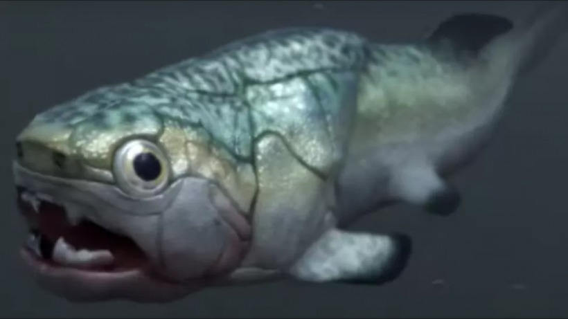
Paleontologists recently discovered a 380-million-year-old heart preserved inside a fossilized prehistoric fish.

The researchers say the specimen captured a key moment in the evolution of the blood-pumping organ found in all back-boned animals, including humans. The heart belonged to a fish known as the Gogo, a prehistoric fish that is now extinct.
The discovery was recently published in the journal Science and was made in Western Australia. Professor Kate Trinajstic, the lead scientist from Curtin University in Perth, Australia, told BBC News about the time when she and her team realized that they had made an unexpected biggest discovery of their lives.

Trinajstic remembered how their team was crowded around the computer and recognized that “they had a heart” and pretty much couldn’t believe it. She uttered that “It was incredibly exciting.

How The Oldest Heart Preserved
The team explained how usually the bones rather than the soft tissues are turned into fossils. The Gogo rock formation minerals have preserved much of the fish’s internal organs, including its stomach, liver, intestine, and the oldest 3D heart.

Trinajstic added that it was a crucial moment in evolution, for the body plan was different in the early centuries compared to the present time because of evolution.
She collaborated with Adelaide’s Flinders University professor John Long. The scientists described the findings as “mind-boggling, jaw-dropping discovery.”
Professor Long said that they had never known anything about the soft internal organs of animals until this latest discovery.

(Photo : Paleozoo) The oldest heart found in the Gogo fish.
Evolution of Gogo Fish’s Heart
The Gogo fish is the first of a class of prehistoric fish that is defined as placoderms. These were the first fish to have physical traits like jaws and teeth. Before the Gogo, fishes were no bigger than 30 centimeters, but the placoderms could grow up to 29.5 feet (9 meters) long.
The placoderms were the earth’s dominant life form for 60 million years, existing for more than 100 million years before the first dinosaurs walked on the planet.
The scans of the Gogo fish fragments showed that its heart was more complex than expected for these primitive fish. The Gogo has two chambers, one on top of the other, similar in structure to the human heart.
The scientists suggest this made the fish’s heart more efficient and became a critical step that transformed it from a slow-moving fish to a fast-paced predator.
Long said that this was the way the Gogo could up the ante and become a voracious predator.

Some other important observation was that the heart was much more forward in the body than those of most primitive fish. The position is believed to have been related to the development of the Gogo fish’s neck and made space for the development of the lungs, and gave further down the evolutionary line.
The Natural History Museum, London Doctor Zerina Johanson, a world leader in placoderms and an independent of Trinajstic’s team, described the study as an “extremely important discovery” that explains why the human body is the way it is in the present time.
It was agreed in the statement of Doctor Martin Brazeau, a placoderm expert from the Imperial College London.
“It’s really exciting to see these results,” Brazeau told BBC News.
This 119 million year old fish, Rhacolepis, is the first fossil to show a 3D preserved heart which gives us a rare window into the early evolution of one of our body’s most important organs. Credit: Dr John Maisey, American Museum of Natural History in New York, Author provided
Palaeontologists and the famous Tin Man in The Wizard of Oz were once in search of the same thing: a heart. But in our case, it was the search for a fossilised heart. And now we’ve found one.
A new discovery, announced today in the journal eLife, shows the perfectly preserved 3D fossilised heart in a 113-119 million-year-old fish from Brazil called Rhacolepis.
This is the first definite fossilised heart found in any prehistoric animal.
For centuries, the fossil remains of back-boned animals – or vertebrates – were studied primarily from their bones or fossilised footprints. The possibility of finding well-preserved soft tissues in really ancient fossils was widely thought to be impossible.
Soft organic material rapidly decays after death, so organs start breaking down from bacterial interactions almost immediately after an animal has died. Once the body has decayed, what remains can eventually become buried and what’s left of the skeleton might one day become a fossil.

Exceptional preservation of fossils
But certain rare fossil deposits, called konservat laggerstätten (meaning “place of storage”), are formed by rapid burial under special chemical conditions. These deposits can preserve a range of soft tissues from the organism.
The fish Rhacolepis imaged by synchrotron tomography showing the heart (left) and a cross-section through the heart showing valves (right, white arrows). Credit: Maldanis et al. (2016)
The famous Burgess Shale fossils from British Columbia in Canada show soft-bodied worms and other invertebrate creatures. These were buried by rapid mudslides around 525 million years ago.
The well-preserved fishes from the 113-119 million-year-old Santana Formation of Brazil were among the first vertebrate fossils to show evidence of preserved soft tissues. These include parts of stomachs and bands of muscles.
The discovery of complete soft tissues preserved as whole internal organs in a fossil was a bit of a Holy Grail for palaeontologists. Such finds could contribute to understanding deeper evolutionary patterns as internal soft organs have their own set of specialised features.

Finding a complete fossilised heart in a fish almost 120 million years old was a major breakthrough for José Xavier-Neto of the Brazilian Biosciences National Laboratory, Lara Maldanis of the University of Campinas, Vincent Fernandez of the European Synchotron Radiation Facility and colleagues from across Brazil and Sweden.
Back in 2000, a group of US scientists claimed to have found a heart preserved in a dinosaur nicknamed Willo, a Thescelosaurus. But recent work has debunked this claim, showing the cavity of the dinosaur body was infilled by sediment and then impregnated with iron-rich minerals to make the cavity inside look a bit heart-like when imaged by CT scanning.
Setting up a fossil in the Australian Synchrotron’s IMBL facility. Fossilised soft organs can be studied using these high-tech imaging methods. Credit: John Long, Flinders University
The only other claims for fossilised vertebrate hearts are stains supposedly made by haemoglobin-rich blood found in the region of the fossil where the heart should be. These, along with stains representing possibly the liver, have recently been documented in 390 million-year-old fishes from Scotland.